Subtypes, lumpectomy versus mastectomy, and the timing of reconstruction.
23.08.2026
The first thing worth understanding is that two women with tumours of identical size can receive entirely different treatment, in a different order, with different drugs. What separates them is the biology of the tumour — the receptors it carries. That result, not the lump, drives the plan.
Pathology testing establishes three things: whether the tumour expresses oestrogen and progesterone receptors, whether it overexpresses HER2, and how fast the cells are dividing.
Hormone receptor positive tumours respond to endocrine therapy — tablets taken for years after the main treatment finishes. HER2 positive tumours, once the worst prognosis in the group, are now treated with targeted antibodies that transformed outcomes for this subtype. Triple negative tumours express none of the three and rely on chemotherapy, with immunotherapy added in some cases.
This is why a treatment plan cannot be proposed from an ultrasound or a mammogram. Anyone offering one has not seen what they need to see.
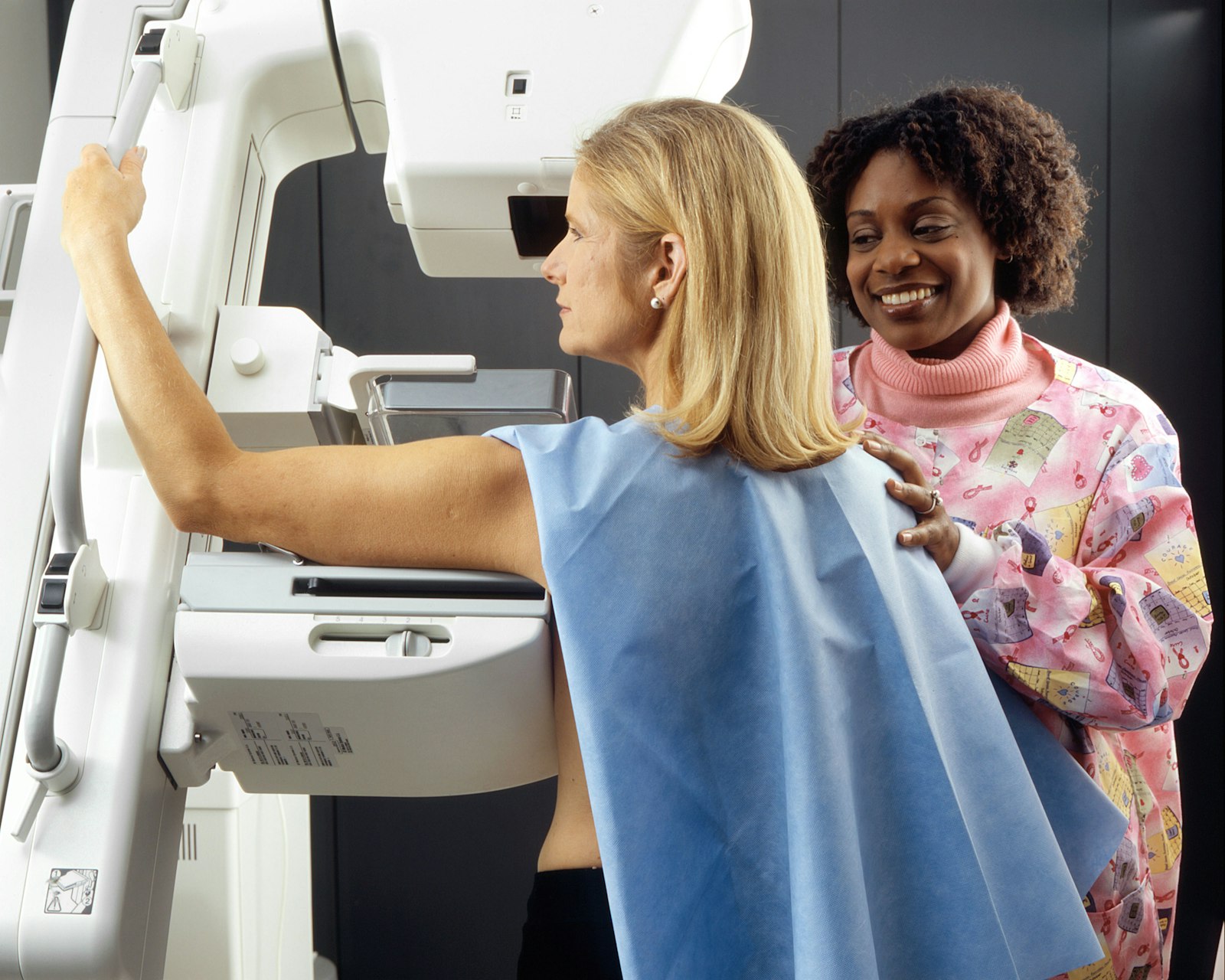
Staging establishes the size of the tumour, whether lymph nodes are involved, and whether disease has spread beyond the breast. This means imaging — mammography, ultrasound, breast MRI where indicated — and assessment of the axillary nodes. Where the tumour is large or the presentation aggressive, PET-CT or other systemic imaging is added.
Genomic testing such as Oncotype DX may be used in hormone positive, node negative cases to estimate whether chemotherapy adds benefit. It is worth asking about, because it can spare some women chemotherapy they would otherwise receive.
Many patients assume the tumour is removed and everything else follows. Often it is the reverse. Neoadjuvant chemotherapy — given before surgery — is standard in HER2 positive and triple negative disease and in larger tumours.
There are two reasons. It can shrink the tumour enough to make breast conserving surgery possible where mastectomy would otherwise be required. And it shows directly whether the tumour responds to the drugs, which is information you cannot obtain any other way. If the tumour has disappeared completely by the time of surgery, that is a meaningful prognostic finding.
Breast conserving surgery removes the tumour with a margin of healthy tissue and is followed by radiotherapy. For appropriately selected patients, survival is equivalent to mastectomy — this is long established and not a compromise.
Mastectomy is indicated where the tumour is large relative to the breast, where there are multiple tumours, where radiotherapy is not possible, or by patient choice. Sentinel lymph node biopsy has largely replaced full axillary clearance where the nodes appear uninvolved, which substantially reduces the risk of lymphoedema.
Immediate reconstruction happens during the same operation as the mastectomy. It avoids a second major surgery and a period without a breast, and the cosmetic result is often better because the skin envelope is preserved. It is not suitable for everyone — planned post-mastectomy radiotherapy complicates it, and some patients are not fit for the longer operation.
Delayed reconstruction happens months or years later, once all treatment is complete. It allows the decision to be made without pressure at the worst possible moment. Techniques divide into implant-based reconstruction and flap reconstruction using your own tissue. Neither is universally better, and the choice should be made with a reconstructive surgeon after all the oncological decisions are settled.
Radiotherapy follows breast conserving surgery essentially always, and follows mastectomy in higher risk cases. Endocrine therapy for hormone positive disease runs five to ten years and is the part patients most often abandon early — side effects are real and worth discussing rather than enduring silently, because there are alternatives within the class.
Follow-up imaging and clinical review continue for years. If you are treated abroad, arrange this follow-up with a doctor at home before you leave, and take a written summary of exactly what was done: the surgery, the pathology, the drugs, the doses, the radiotherapy field and dose.
Can I have reconstruction at the same time as the mastectomy?
Often yes, and immediate reconstruction avoids a second major operation. Whether it is appropriate depends on the tumour, on whether radiotherapy is planned, and on your general fitness.
Is a lumpectomy less effective than a mastectomy?
No. For appropriately selected patients, breast conserving surgery with radiotherapy gives equivalent survival. The choice depends on tumour size and position relative to the breast.
What do you need to review my case?
The pathology report including receptor status and HER2, all imaging on disc, and any treatment already given. Receptor status is the single most important document.
Related reading
CyberKnife, Gamma Knife and Proton Therapy: What Turkey Actually Has
Getting a Second Opinion on a Cancer Diagnosis from Another Country
How Quickly Can Cancer Treatment Start in Turkey? A Realistic Timeline
This page is general information, not medical advice. Diagnosis, surgical options, drug therapy and reconstruction are determined individually by a licensed medical team.
Send us what you already have — the pathology report, imaging on disc, discharge summaries. A specialist at a partner hospital will review it and tell you what is realistic in your case and how quickly treatment could begin. Reviewing your documents costs nothing and commits you to nothing.
Send your documentsUnhappy with a surgery result abroad? How to tell unfinished healing from a real problem, what a revision involves, who pays, and where to go next.
Silicone oil removal after vitrectomy: why the oil is used, how long it stays, how the second operation works and what vision does afterwards.
How children treatment in Turkey actually works: the accompanying parent, documents for a minor, school, language, paediatric specialties and what to send.